Save that Tooth! Alternatives to “Toothanasia”
For Pet Owners

Most veterinarians know that teeth with infected pulp or advanced periodontal disease need to be treated, but many are not aware that there are alternatives to tooth extraction. Veterinary dental specialists can perform procedures to save endontically diseased or periodontally compromised teeth.
Endodontically diseased teeth can often be saved with vital pulp therapy or root canal treatment. These procedures, in most cases, allow broken or non-vital teeth to remain in pets’ mouths for the rest of their lives.
Fractured teeth with exposed pulp are painful and inevitably become infected. Studies have also shown that more than 90% of discolored teeth in dogs are dead and have infected pulp. Extraction of these diseased teeth removes the risk of pain and infection, but the pet no longer has use of these teeth.
Endodontic treatment—vital pulp therapy or root canal therapy—are preferred alternatives to extraction, particularly for important teeth like canine and carnassial teeth in dogs. Endodontic treatment is also an excellent option for canine teeth in cats, but is more difficult in other feline teeth because of the small size of the pulp canals.
Vital pulp therapy, also called vital pulpotomy, can be performed on fractured teeth with pulp exposure where exposure has occurred within 48 hours of treatment. In this procedure, a small amount of infected pulp is removed and a dressing powder called mineral trioxide aggregate (MTA) is placed on the pulp. The fracture site is sealed using composite--the same material used for filling cavities in humans. The dressing stimulates the tooth to form a bridge of dentin over the pulp and the tooth usually remains alive and healthy.
Root canal treatment on teeth with older fractures or on non-vital teeth can also be performed. The infected pulp and a thin layer of dentin lining the pulp canal is removed from the tooth using endodontic files. The pulp canal is flushed with disinfectant and the tooth is filled with an inert sealant called gutta percha. The fracture site and site used to access the pulp canal are filled with composite.
Both of these procedures have a better than 90% success rate in dogs and most likely have a similar success rate in cats, although this has not been studied. Dental radiographs of treated teeth should be taken annually to make sure the treatment has been successful.
Periodontal disease starts with accumulation of plaque bacteria, which hardens into calculus (tartar). This leads to the earliest form of periodontal disease—inflamed gums (gingivitis). Gingivitis can be reversed with a professional cleaning under anesthesia to remove the calculus and daily home care, including tooth-brushing. If untreated, periodontal disease can progress to bone loss, loose teeth and oral infection.
Teeth with moderate to advanced periodontal disease do not always need to be extracted. These teeth can often be treated using advanced periodontal techniques. Open root planing is performed by creating a gingival flap and removing plaque bacteria and calculus from periodontal pockets. This regenerates attachment of the gums and can save teeth with moderate periodontal disease. Sometimes a long-acting antibiotic, or perioceutical is placed in the pocket to help encourage gingival reattachment.
Teeth with deep pockets can often be saved by performing open root planing, followed by guided tissue regeneration to replace lost supporting bone. In guided tissue regeneration, bone-grafting material is placed in periodontal pockets and a resorbable collagen membrane is placed over the graft. The membrane prevents soft tissue from migrating into the pocket until new periodontal ligament and bone regenerates. These treatments are only suitable for some types of periodontal bone loss and a veterinary dental specialist needs to evaluate teeth with radiographs and an oral examination under anesthesia in order to determine if they are candidates for these procedures.
Figures
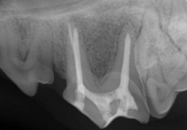
Figure 1: Fourth premolar tooth after root canal treatment
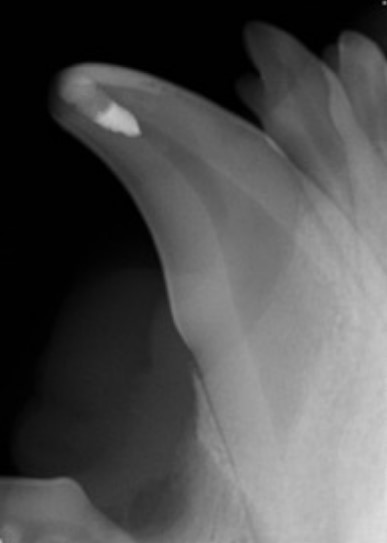
Figure 2: Canine tooth immediately after vital pulp therapy
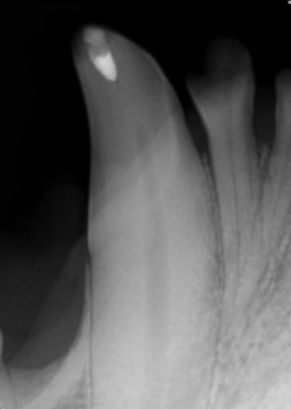
Figure 3: Canine tooth six months after vital pulp therapy. A dental bridge has formed just below the dressing material at the fracture site and the pulp canal has narrowed indicating the tooth is vital and healthy
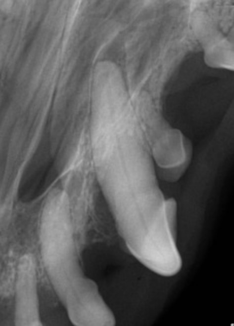
Figure 4: Canine tooth with a deep periodontal pocket before guided tissue regeneration \
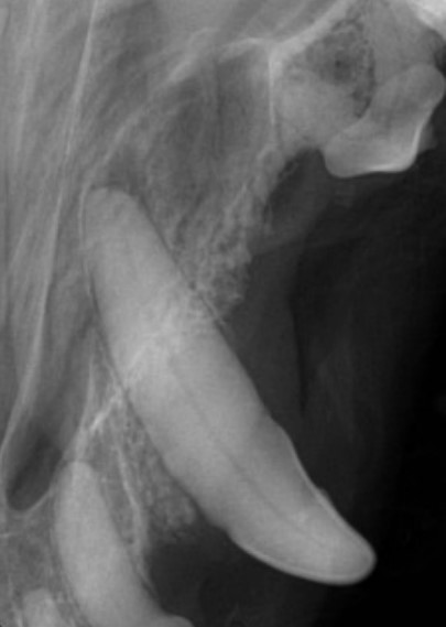
Figure 5: Canine tooth after guided tissue regeneration. The periodontal pocket has been populated with bone and a new periodontal ligament space is present indicating regeneration of the periodontal ligament. (First premolar tooth has been extracted)
